
12.2: PNS - Nerves and Ganglia
- Page ID
- 64063
The PNS is not as contained as the CNS because it is defined as everything that is not the CNS. Some peripheral structures are incorporated into the other organs of the body. In describing the anatomy of the PNS, it is necessary to describe the common structures, the nerves and the ganglia, as they are found in various parts of the body. Many of the neural structures that are incorporated into other organs are features of the digestive system; these structures are known as the enteric nervous system and are a special subset of the PNS.
Nerve Structure
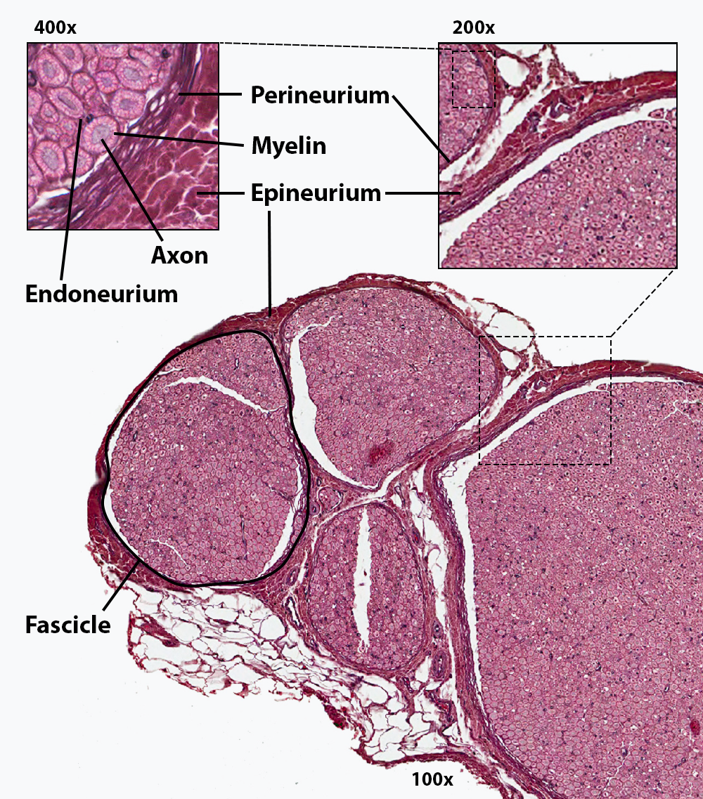
Bundles of axons in the PNS are referred to as nerves. These structures in the periphery are different than their central counterpart, called a tract. Nerves are organs, so they are composed of more than just nervous tissue. They have connective tissues invested in their structure, as well as blood vessels supplying the tissues with nourishment. The outer surface of a nerve is a surrounding layer of dense irregular connective tissue called the epineurium. Within the nerve, axons are further bundled into fascicles, which are each surrounded by their own layer of dense irregular connective tissue called the perineurium. Finally, individual axons are surrounded by areolar connective tissue called the endoneurium (Figure \(\PageIndex{1}\). These three layers are similar to the connective tissue sheaths for muscles. Nerves are associated with the region of the CNS to which they are connected, either as cranial nerves connected to the brain or spinal nerves connected to the spinal cord.
Ganglia
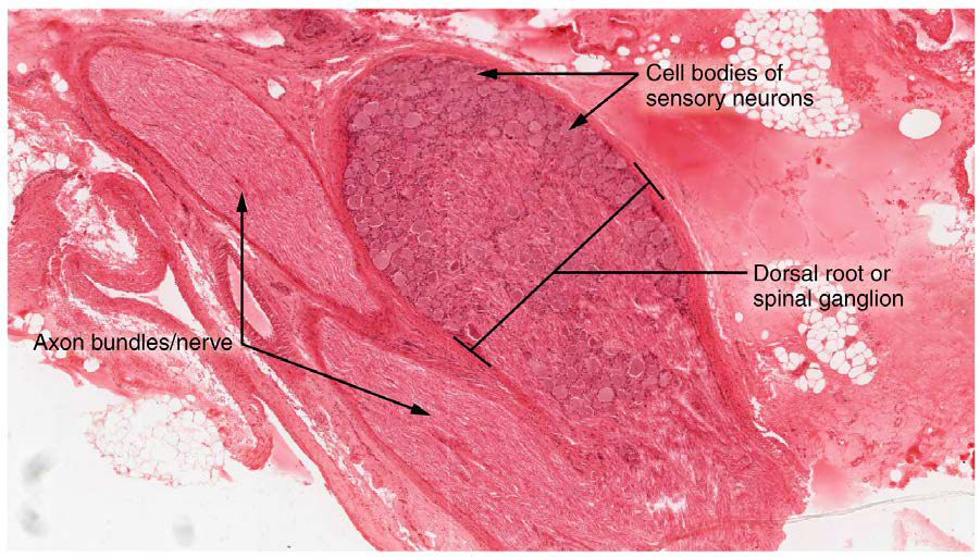
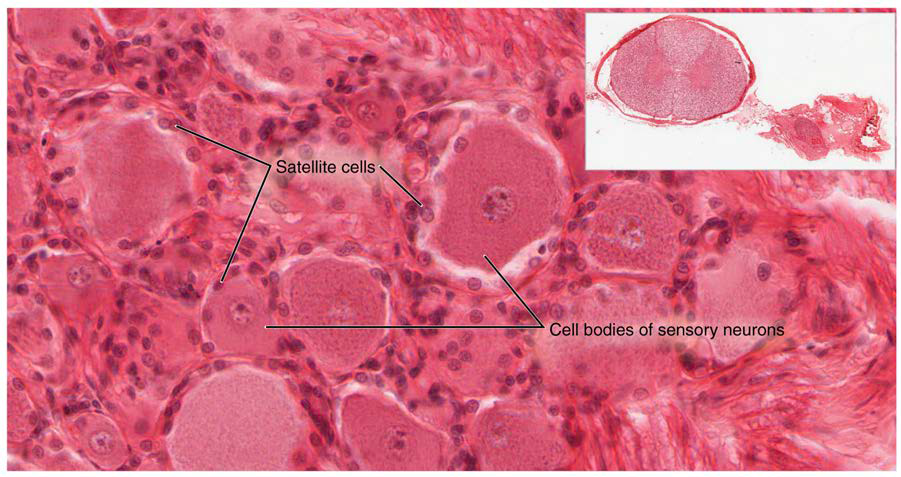
A ganglion (ganglia for plural) is a group of neuron cell bodies in the peripheral nervous system. Ganglia can be categorized, for the most part, as either sensory ganglia or autonomic ganglia, referring to their primary functions. A type of sensory ganglion is a cranial nerve ganglion. The roots of cranial nerves are within the cranium, whereas the ganglia are outside the skull. For example, the trigeminal ganglion is superficial to the temporal bone whereas its associated nerve is attached to the mid-pons region of the brainstem. The neurons of cranial nerve ganglia are also unipolar in shape with associated satellite cells. Another type of sensory ganglion is found associated with the spinal nerves - the posterior (dorsal) root ganglia (Figure \(\PageIndex{2}\)). These ganglia, one for each spinal nerve, house the cell bodies of the sensory neurons bringing sensation from somatic regions inferior to the cranium. Within the spinal ganglia, the cell bodies of the unipolar neurons are surrounded by satellite cells (Figure \(\PageIndex{3}\)).
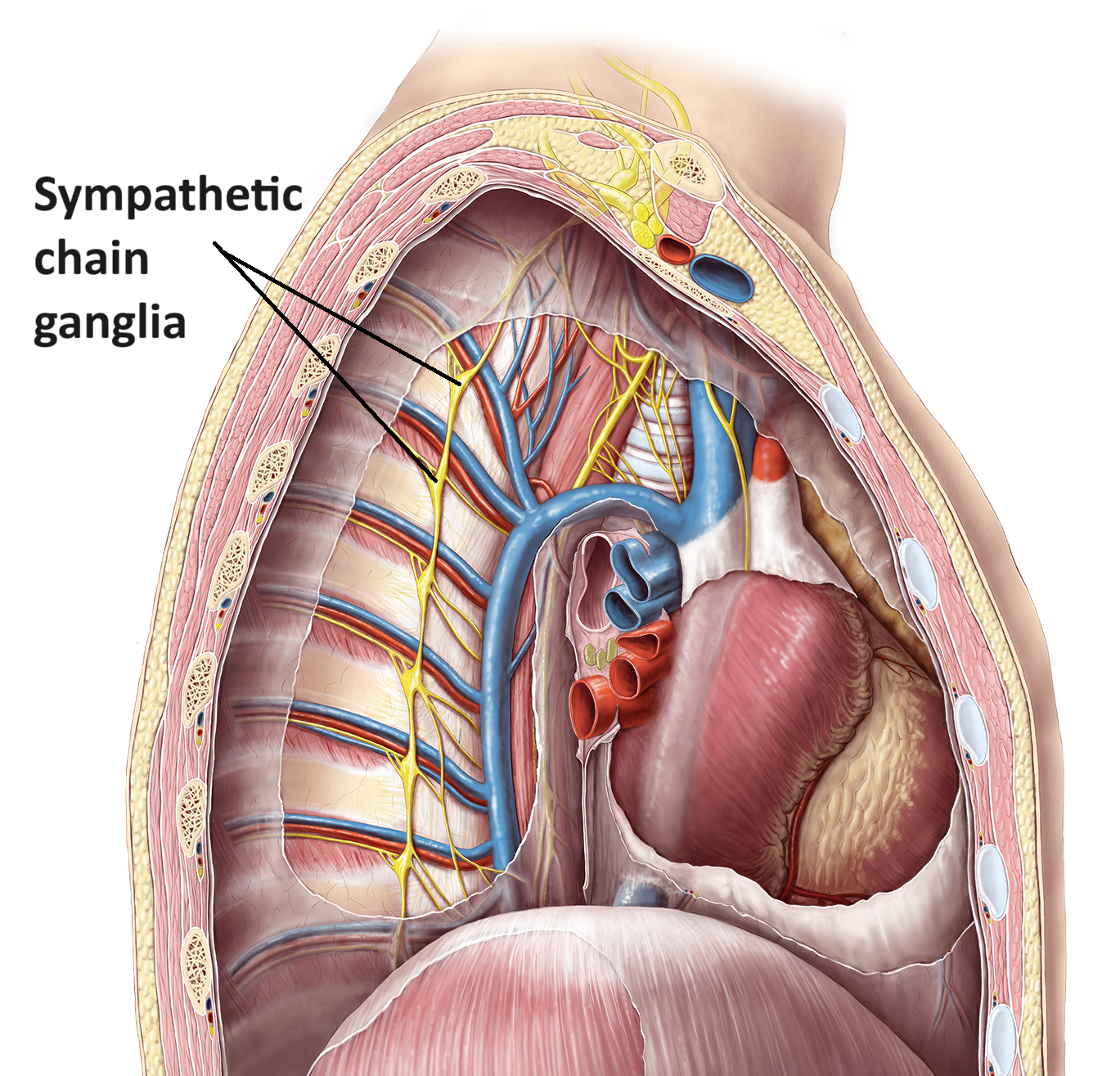
Autonomic ganglia differ in structure from the sensory ganglia because they contain the synapse point for the preganglionic cells projecting from the lateral horns of the spinal cord. In response to incoming signals, the neurons in the autonomic ganglia generate a new action potential that will be carried along the postganglionic axon to the effector organ/tissue. Examples of autonomic ganglia are the sympathetic chain ganglia (Figure \(\PageIndex{4}\)) located lateral to the vertebral column and the otic ganglion near the foramen ovale.
Concept Review
The PNS is composed of the groups of neurons (ganglia) and bundles of axons (nerves) that are outside of the brain and spinal cord. Nerves are organized into structures by layers of connective tissue that cover them. The epineurium covers the nerve, the perineurium covers the fascicles and the endoneurium covers the individual axon.
Nerves are classified as cranial nerves or spinal nerves on the basis of their connection to the brain or spinal cord, respectively.
Ganglia are of two types, sensory or autonomic. Sensory ganglia contain unipolar sensory neurons and are associated with many of the cranial nerves.
Review Questions
Q. What type of ganglion contains neurons that control autonomic mechanisms of the body?
A. sensory ganglion
B. dorsal root ganglion
C. sympathetic chain ganglion
D. ventral root ganglion
- Answer
-
C
Q. What is the name for a bundle of axons within a nerve?
A. fascicle
B. tract
C. nerve root
D. epineurium
- Answer
-
A
Q. The endoneurium surrounds _______.
A. a nerve
B. a bundle of fascicles
C. a bundle of myelinated axons
D. a myelinated axon
- Answer
-
D
Critical Thinking Questions
Q. Why are ganglia and nerves not surrounded by protective structures like the meninges of the CNS?
A. The peripheral nervous tissues are out in the body, sometimes part of other organ systems. There is not a privileged blood supply like there is to the brain and spinal cord, so peripheral nervous tissues do not need the same sort of protections.
Glossary
- cranial nerve ganglion
- sensory ganglion of cranial nerves
- endoneurium
- innermost layer of connective tissue that surrounds individual axons within a nerve
- epineurium
- outermost layer of connective tissue that surrounds an entire nerve
- fascicle
- small bundles of nerve or muscle fibers enclosed by connective tissue
- perineurium
- layer of connective tissue surrounding fascicles within a nerve
- trigeminal ganglion
- sensory ganglion that contributes sensory fibers to the trigeminal nerve
Contributors and Attributions
OpenStax Anatomy & Physiology (CC BY 4.0). Access for free at https://openstax.org/books/anatomy-and-physiology