
12.3: Cranial Nerves
- Page ID
- 63444
- Name the twelve cranial nerves and explain the functions associated with each
Cranial Nerves
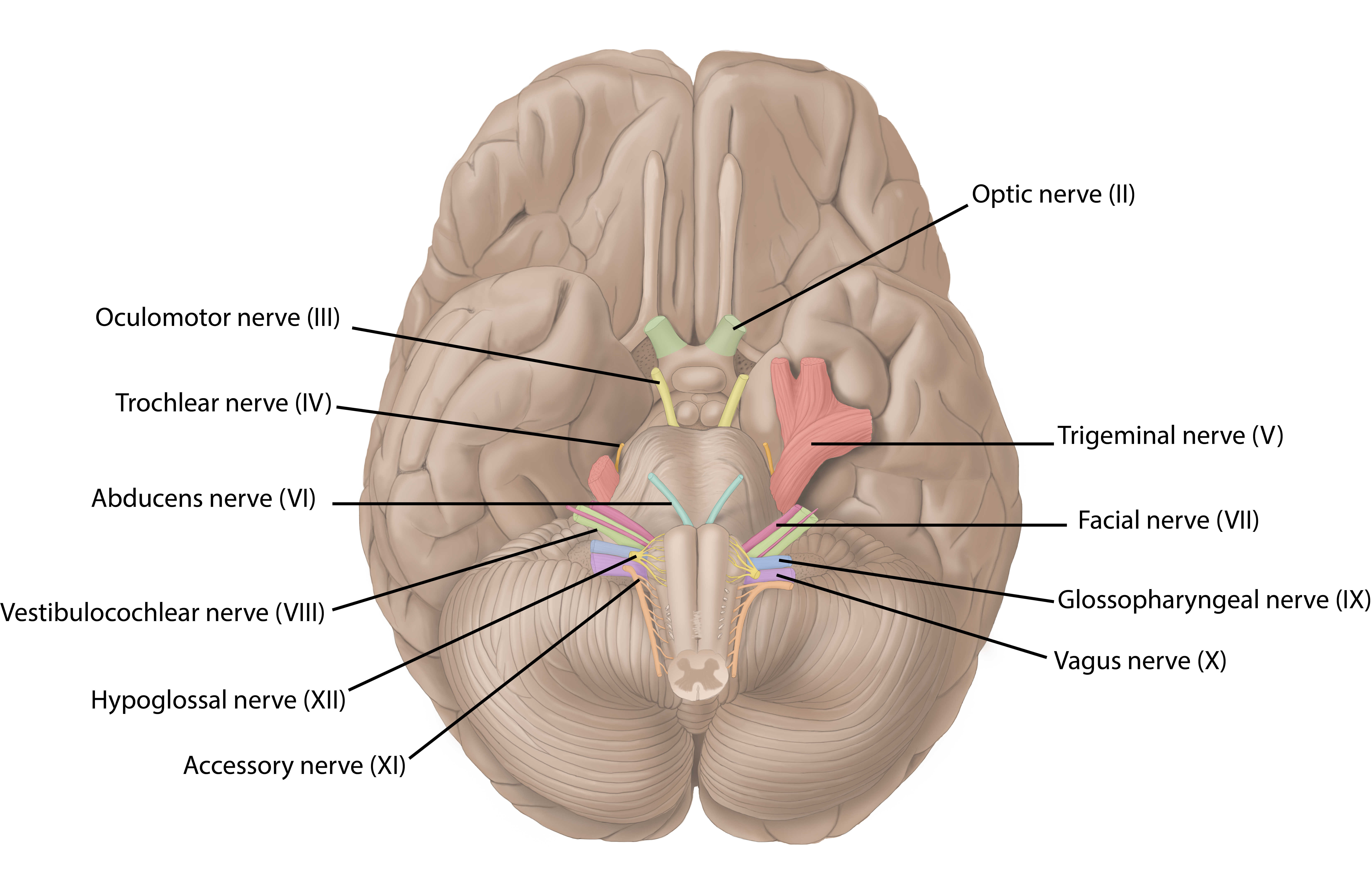
The nerves attached to the brain are the cranial nerves, which are primarily responsible for the sensory and motor functions of the head and neck (with the exception of one that targets organs in the thoracic and abdominal cavities as part of the parasympathetic nervous system). There are twelve cranial nerves, which are designated CN I through CN XII for “Cranial Nerve,” using Roman numerals for 1 through 12, based on the anatomical location on the inferior view of the brain, from anterior to posterior (Figure \(\PageIndex{1}\)). They can be classified as sensory nerves, motor nerves, or a combination of both, meaning that the axons in these nerves originate out of sensory ganglia external to the cranium or motor nuclei within the brainstem. Sensory axons enter the brain to synapse in a nucleus. Motor axons connect to skeletal muscles of the head or neck. Three of the nerves are solely composed of sensory fibers; five are strictly motor; and the remaining four are mixed nerves.
Learning the cranial nerves is a tradition in anatomy courses, and students have always used mnemonic devices to remember the nerve names. A traditional mnemonic is the rhyming couplet, “Oh Oh Oh To Touch And Feel Very Good Velvet Ah Heaven,” in which the initial letter of each word corresponds to the initial letter in the name of each nerve. The names of the nerves have changed over the years to reflect current usage and more accurate naming. An exercise to help learn this sort of information is to generate a mnemonic using words that have personal significance. The names of the cranial nerves are listed in Table \(\PageIndex{1}\) along with a brief description of their function, their source (sensory ganglion or motor nucleus), and their target (sensory nucleus or skeletal muscle).
- The olfactory nerve is responsible for the sense of smell. It is usually not seen on the brain as its short fibers pass through the cribriform plate and thus are torn when the brain is extracted. Instead, we see the afferent nerves: the olfactory bulb and the olfactory tract.
- The optic nerves are responsible for the sense of vision.
- The oculomotor nerve is responsible for eye movements by controlling four of the extraocular muscles. It is also responsible for lifting the upper eyelid when the eyes point up, for pupillary constriction and focusing light by adjusting the lens of the eye..
- The trochlear nerve and the abducens nerve are both responsible for eye movement, each controlling a singular extraocular muscle.
- The trigeminal nerve is responsible for cutaneous sensations of the face and mouth and controlling the muscles of mastication. It also has a small contribution to the sensation of taste.
- The facial nerve is responsible for the muscles involved in facial expressions. Its sensory components include part of the sense of taste, the production of tears, and the production of saliva.
- The vestibulocochlear nerve is responsible for the senses of hearing and balance.
- The glossopharyngeal nerve is responsible for controlling muscles in the oral cavity and upper throat and the production of saliva. Its sensory components include taste from the posterior tongue, blood pressure, and blood pH, pO2, and pCO2.
- The vagus nerve is responsible for both sensory and motor from/to the organs of the thoracic and abdominal cavities. It also carries motor signals to the muscles of the larynx, pharynx and soft palate. Additional sensory functions include taste and general sensory from the larynx. In innervating this broad area, the vagus nerve is responsible for the regulation of internal organ functions, such as digestion, heart rate, and respiratory rate, as well as vasomotor activity, and certain reflex actions, such as coughing, sneezing, swallowing, and vomiting.
- The accessory nerve's spinal component is responsible for controlling two muscles of the neck, the sternocleidomastoid and the superior portion of the trapezius.
- The hypoglossal nerve is responsible for controlling the muscles of the tongue.
Another important aspect of the cranial nerves that lends itself to a mnemonic is the functional role each nerve plays. The nerves fall into one of three basic groups. They are sensory, motor, or both (see Table \(\PageIndex{1}\)). The sentence, “Some Say Marry Money But My Brother Says Brains Beauty Matter More,” corresponds to the type(s) of information carried by each nerve:
- Sensory only: the first, second, and eighth nerves are purely sensory: the olfactory (CN I), optic (CN II), and vestibulocochlear (CN VIII) nerves.
- Motor only: The three eye-movement nerves are all only motor: the oculomotor (CN III), trochlear (CN IV), and abducens (CN VI). The accessory (CN XI) and hypoglossal (CNXII) nerves are also strictly motor.
- Both: The remainder of the nerves contain both sensory and motor fibers, these are mixed nerves: the trigeminal (CN V), facial (CN VII), glossopharyngeal (CN IX), and vagus (CN X) nerves.
| Mnemonic | # | Name | Function (S/M/B) | Central connection (nuclei) | Peripheral connection (ganglion or muscle) |
|---|---|---|---|---|---|
| Oh | I | Olfactory | Smell (S) | Olfactory bulb | Olfactory epithelium |
| Oh | II | Optic | Vision (S) | Hypothalamus/thalamus/midbrain | Retina (retinal ganglion cells) |
| Oh | III | Oculomotor | Eye movements (M) | Oculomotor nucleus | Extraocular muscles (other 4), levator palpebrae superioris, ciliary ganglion (autonomic) |
| To | IV | Trochlear | Eye movements (M) | Trochlear nucleus | Superior oblique muscle |
| Touch | V | Trigeminal | Sensory/motor – face (B) | Trigeminal nuclei in the midbrain, pons, and medulla | Trigeminal |
| And | VI | Abducens | Eye movements (M) | Abducens nucleus | Lateral rectus muscle |
| Feel | VII | Facial | Motor – face, Taste (B) | Facial nucleus, solitary nucleus, superior salivatory nucleus | Facial muscles, Geniculate ganglion, Pterygopalatine ganglion (autonomic) |
| Very | VIII | Vestibulocochlear | Hearing/balance (S) | Cochlear nucleus, Vestibular nucleus/cerebellum | Spiral ganglion (hearing), Vestibular ganglion (balance) |
| Good | IX | Glossopharyngeal | Motor – throat Taste (B) | Solitary nucleus, inferior salivatory nucleus, nucleus ambiguus | Pharyngeal muscles, Geniculate ganglion, Otic ganglion (autonomic) |
| Velvet | X | Vagus | Motor/sensory – viscera (autonomic) (B) | Medulla | Terminal ganglia serving thoracic and upper abdominal organs (heart and small intestines) |
| Ah | XI | Accessory | Motor – head and neck (M) | Spinal accessory nucleus | Neck muscles |
| Heaven | XII | Hypoglossal | Motor – lower throat (M) | Hypoglossal nucleus | Muscles of the larynx and lower pharynx |
Vision Loss

Read this article about a man who wakes with a headache and a loss of vision. His regular doctor sent him to an ophthalmologist to address the vision loss. The ophthalmologist recognizes a greater problem and immediately sends him to the emergency room. Once there, the patient undergoes a large battery of tests, but a definite cause cannot be found. A specialist recognizes the problem as meningitis, but the question is what caused it originally. How can that be cured? The loss of vision comes from swelling around the optic nerve, which probably presented as a bulge on the inside of the eye. Why is swelling related to meningitis going to push on the optic nerve?
- Answer
-
The optic nerve enters the CNS in its projection from the eyes in the periphery, which means that it crosses through the meninges. Meningitis will include swelling of those protective layers of the CNS, resulting in pressure on the optic nerve, which can compromise vision.
Nervous System: Anosmia
Anosmia is the loss of the sense of smell. It is often the result of the olfactory nerve being severed, usually because of blunt force trauma to the head. The sensory neurons of the olfactory epithelium have a limited lifespan of approximately one to four months, and new ones are made on a regular basis. The new neurons extend their axons into the CNS by growing along the existing fibers of the olfactory nerve. The ability of these neurons to be replaced is lost with age. Age-related anosmia is not the result of impact trauma to the head, but rather a slow loss of the sensory neurons with no new neurons born to replace them.
Smell is an important sense, especially for the enjoyment of food. There are only five tastes sensed by the tongue, and two of them are generally thought of as unpleasant tastes (sour and bitter). The rich sensory experience of food is the result of odor molecules associated with the food, both as food is moved into the mouth, and therefore passes under the nose, and when it is chewed and molecules are released to move up the pharynx into the posterior nasal cavity. Anosmia results in a loss of the enjoyment of food.
As the replacement of olfactory neurons declines with age, anosmia can set in. Without the sense of smell, many sufferers complain of food tasting bland. Often, the only way to enjoy food is to add seasoning that can be sensed on the tongue, which usually means adding table salt. The problem with this solution, however, is that this increases sodium intake, which can lead to cardiovascular problems through water retention and the associated increase in blood pressure.
Autonomic Functions
Four of these cranial nerves make up the cranial component of the autonomic nervous system, as they innervate smooth muscle, cardiac muscle, or glands.
- The oculomotor nerves are responsible for innervating the intra-ocular muscles responsible for pupillary diameter and adjusting the eyes' focal point.
- The facial and glossopharyngeal nerves control salivation and lacrimation, which are glandular secretions.
- The regulation of the organs of the thoracic and abdominal cavities such as the heart, bronchial tree, stomach, pancreas, adrenal glands, and kidneys.
Concept Review
The twelve cranial nerves can be strictly sensory in function, strictly motor in function, or a combination of the two functions. The olfactory nerve (CN I) and optic nerve (CN II) are responsible for the sense of smell and vision, respectively. The oculomotor nerve (CN III) is responsible for eye movements, lifting the upper eyelid and size of the pupil. The trochlear nerve (CN IV) and the abducens nerve (CN VI) are both responsible for eye movement, but do so by controlling different extraocular muscles. The trigeminal nerve (CN V) is responsible for cutaneous sensations of the face and controlling the muscles of mastication. The facial nerve (VII) is responsible for the muscles involved in facial expressions, as well as part of the sense of taste and the production of saliva. The vestibulocochlear nerve (VIII) is responsible for the senses of hearing and balance. The glossopharyngeal nerve (IX) is responsible for controlling muscles in the oral cavity and upper throat, as well as part of the sense of taste and the production of saliva. The vagus nerve (CN X) is responsible for contributing to homeostatic control of the organs of the thoracic and upper abdominal cavities. The accessory nerve (CN XI) is responsible for controlling the muscles of the neck, along with cervical spinal nerves. The hypoglossal nerve (CN XII) is responsible for controlling the muscles of the lower throat and tongue.
Review Questions
Q. How many cranial nerves carry sensory information for taste?
A. 1
B. 2
C. 3
D. 4
- Answer
-
D
Q. Which cranial nerve does not control organs in the head and neck?
A. olfactory
B. trochlear
C. glossopharyngeal
D. vagus
- Answer
-
D
Q. Which cranial nerve has both somatic and visceral motor functions?
A. oculomotor
B. trochlear
C. trigeminal
D. hypoglossal
- Answer
-
A
Critical Thinking Questions
Q. If a patient's pupils do no respond properly when a bright light is shown into them, which cranial nerves need to be checked to make sure they are functioning?
A. The pupillary light reflex causes the pupils to dilate in low light conditions and constrict in high light conditions. If a patient's eyes fail to make these adjustments then both a sensory and a motor defect need to be considered. Can the patient see the light? Do the muscles controlling pupil diameter contract and relax? Sensory information related to light is carried by the optic nerves (CN II) and motor control to the muscles of the the pupils is carried by the oculomotor nerves (CN III), so both of these need to be considered.
Q. Testing for neurological function involves a series of tests of functions associated with the cranial nerves. What functions, and therefore which nerves, are being tested by asking a patient to follow the tip of a pen with their eyes?
A. The contraction of extraocular muscles is being tested, which is the function of the oculomotor, trochlear, and abducens nerves.
Glossary
- abducens nerve
- sixth cranial nerve; responsible for contraction of one of the extraocular muscles
- accessory nerve
- eleventh cranial nerve; responsible for contraction of neck muscles
- cranial nerve
- one of twelve nerves connected to the brain that are responsible for sensory or motor functions of the head and neck
- facial nerve
- seventh cranial nerve; responsible for contraction of the facial muscles and for part of the sense of taste, as well as causing saliva production
- glossopharyngeal nerve
- ninth cranial nerve; responsible for contraction of muscles in the tongue and throat and for part of the sense of taste, as well as causing saliva production
- hypoglossal nerve
- twelfth cranial nerve; responsible for contraction of muscles of the tongue
- oculomotor nerve
- third cranial nerve; responsible for contraction of four of the extraocular muscles, the muscle in the upper eyelid, and pupillary constriction
- olfactory nerve
- first cranial nerve; responsible for the sense of smell
- optic nerve
- second cranial nerve; responsible for visual sensation
- trigeminal nerve
- fifth cranial nerve; responsible for cutaneous sensation of the face and contraction of the muscles of mastication
- trochlear nerve
- fourth cranial nerve; responsible for contraction of one of the extraocular muscles
- vagus nerve
- tenth cranial nerve; responsible for the autonomic control of organs in the thoracic and upper abdominal cavities
- vestibulocochlear nerve
- eighth cranial nerve; responsible for the sensations of hearing and balance
Contributors and Attributions
OpenStax Anatomy & Physiology (CC BY 4.0). Access for free at https://openstax.org/books/anatomy-and-physiology