
12.4: Spinal Nerves and Their Branches
- Page ID
- 63445
- Describe the sensory and motor components of spinal nerves and the plexuses that they pass through
- Explain what a dermatome is and its clinical significance
Along the spinal cord, spinal nerves emerge that carry both sensory and motor information.
Spinal Nerves
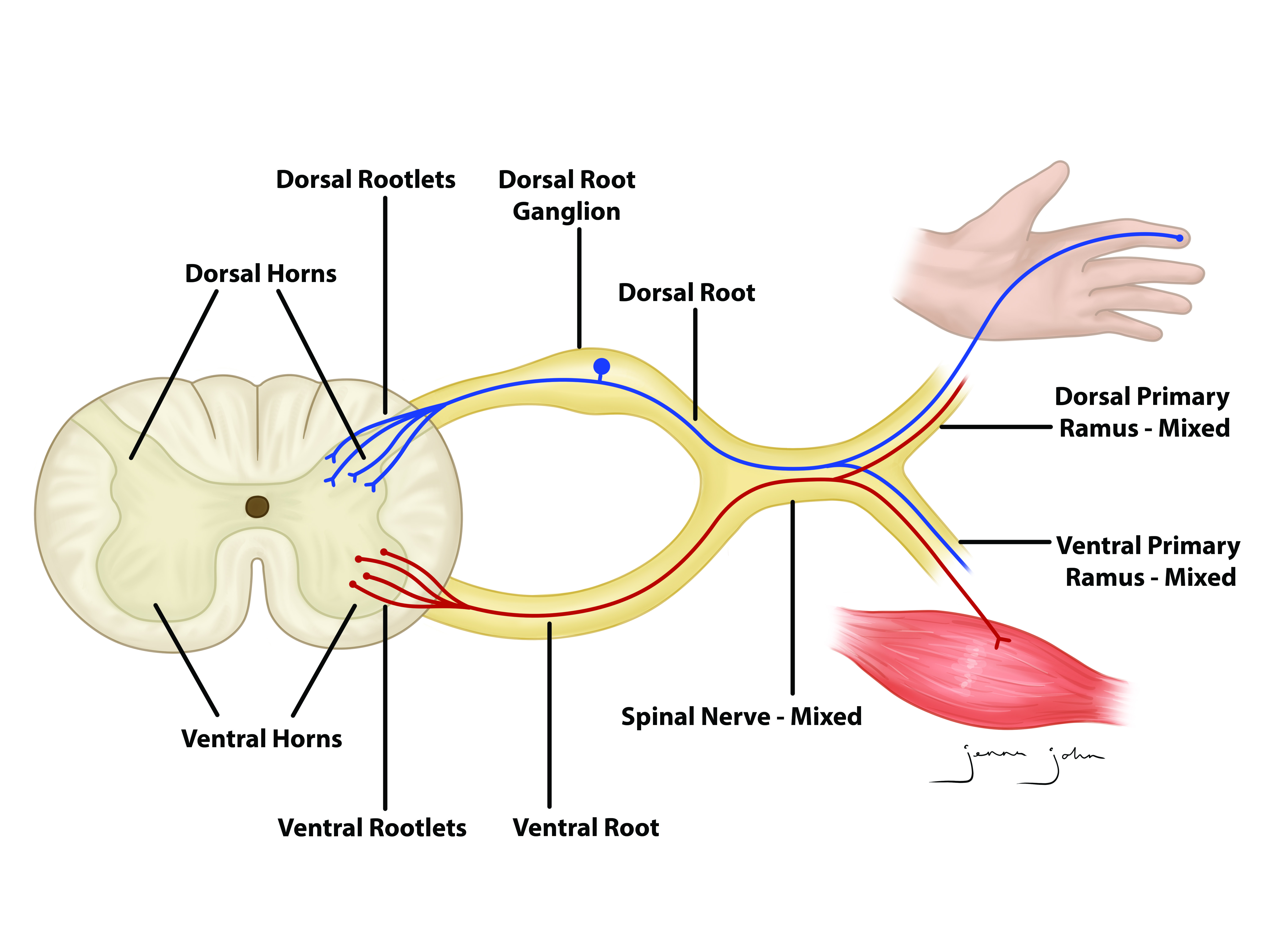
The nerves connected to the spinal cord are the spinal nerves. The arrangement of these nerves is much more regular than that of the cranial nerves. All of the spinal nerves are combined sensory and motor axons that separate into two nerve roots. The sensory axons enter the spinal cord as the dorsal nerve root. The motor fibers, both somatic and autonomic, emerge as the ventral nerve root.
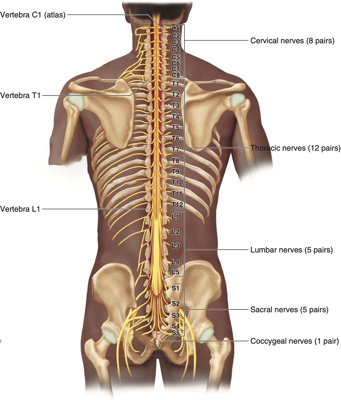
There are 31 spinal nerves, named for the level of the spinal cord at which each one emerges (Figure \(\PageIndex{1}\)). There are
- eight pairs of cervical nerves designated C1 to C8,
- twelve pairs of thoracic nerves designated T1 to T12,
- five pairs of lumbar nerves designated L1 to L5,
- five pairs of sacral nerves designated S1 to S5, and
- one pair of coccygeal nerves designated Co1.
The nerves are numbered from the superior to inferior positions, and each emerges from the vertebral column through the intervertebral foramen at its level. The first nerve, C1, emerges between the first cervical vertebra and the occipital bone. The second nerve, C2, emerges between the first and second cervical vertebrae. The same occurs for C3 to C7, but C8 emerges between the seventh cervical vertebra and the first thoracic vertebra. For the thoracic and lumbar nerves, each one emerges between the vertebra that has the same designation and the next vertebra in the column. The sacral nerves emerge from the sacral foramina along the length of that unique vertebra.
Spinal Nerve Branching
As shown in Figure \(\PageIndex{2}\), axons coming from the posterior (dorsal) root ganglion enter the posterior side through the posterior (dorsal) nerve root. The axons emerging from the anterior side do so through the anterior (ventral) nerve root. The posterior and anterior nerve roots fuse together to form the spinal nerves.
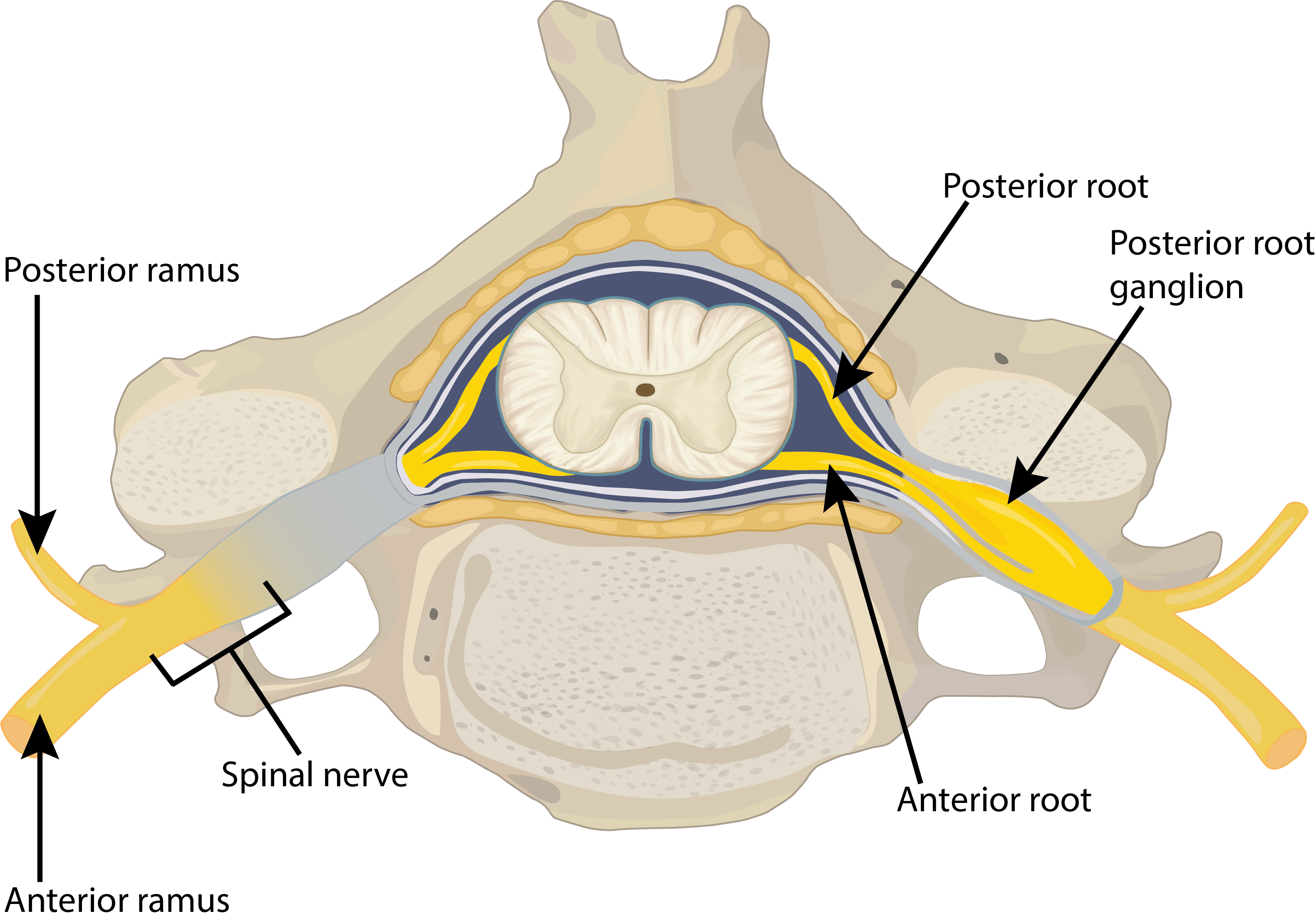
The posterior and anterior nerve roots join to form the spinal nerve, which thus contains mixed sensory and motor information. The spinal nerves are quite short. Lateral to the vertebral column the nerve fibers split and head to different locations in the body via the rami: those traveling in the posterior (dorsal) ramus serve the muscles and skin of the posterior trunk, those in the larger anterior (ventral) ramus travel either to the anterolateral trunk or to the limbs. Both the anterior and posterior rami are mixed nerves carrying somatic motor fibers and sensory fibers. The visceral motor fibers take a different path via the rami communicans (grey and white rami), which will be explored more in Chapter 14.
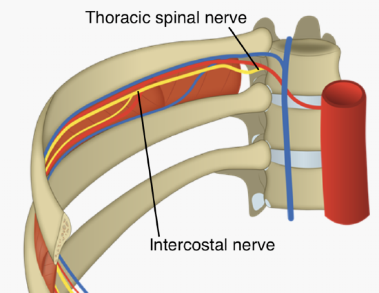
In the thoracic region (T2-T12), the ventral rami are called intercostal nerves because they travel deep to the intercostal muscles in the space between adjacent ribs (Figure \(\PageIndex{4}\)).
Nerve Plexuses
In the cervical, lumbar and sacral regions the nerves in the periphery are not straight continuations of the spinal nerves, but rather the reorganization of the axons in those nerves to follow different courses. Axons from different spinal nerves merge and split multiple times at four places along the length of the vertebral column, each identified as a nerve plexus (plexus = a braid). Of the four nerve plexuses, two are found at the cervical level, one at the lumbar level, and one at the sacral level (Figure \(\PageIndex{5}\)).
The cervical plexus is composed of axons from spinal nerves C1 through C5. It branches into nerves in the posterior neck and head as well as the phrenic nerve, which connects to the diaphragm at the base of the thoracic cavity.
.png?revision=1&size=bestfit&width=625&height=733)
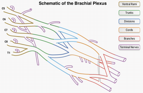
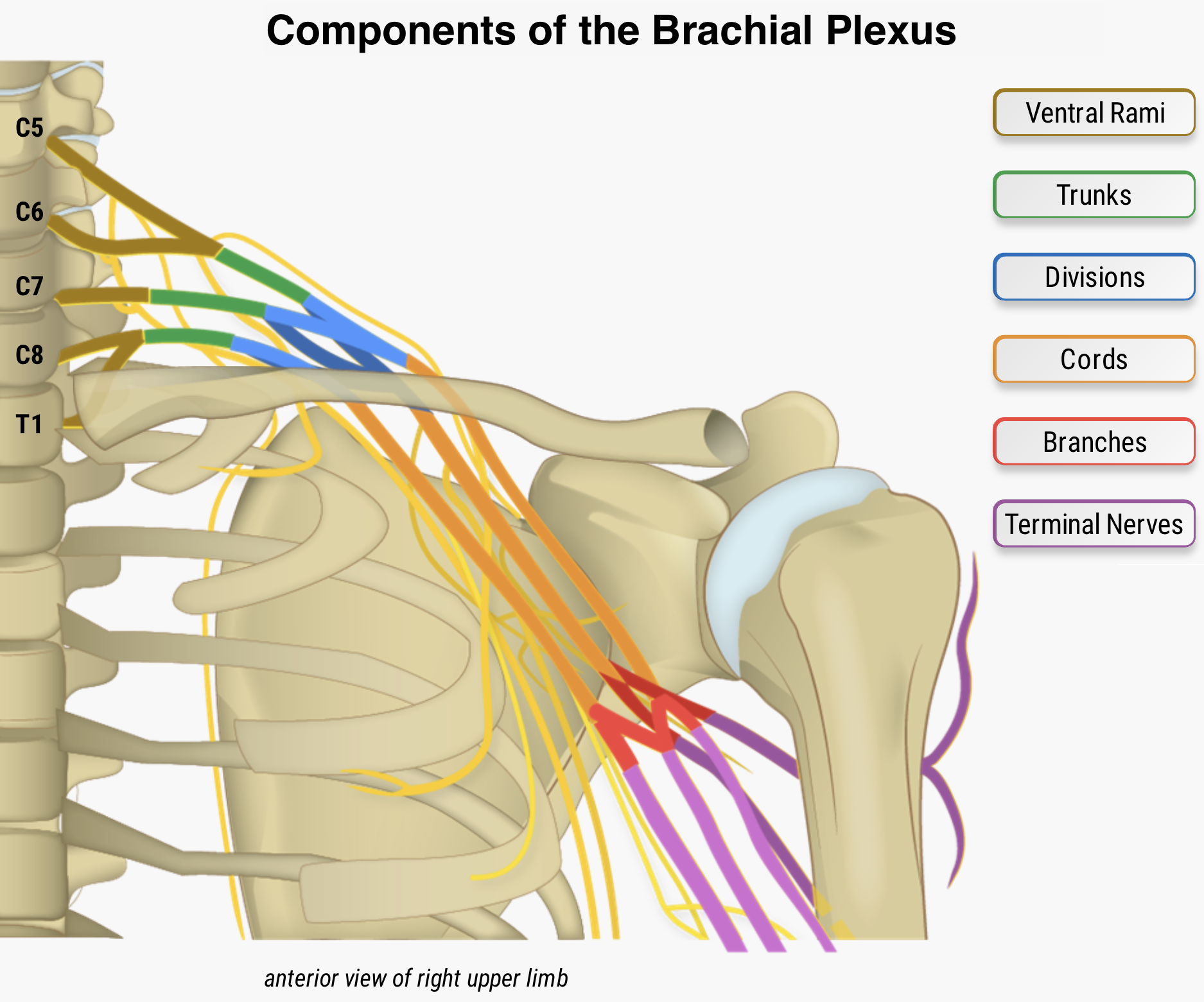
The other plexus from the cervical level is the brachial plexus (Figure \(\PageIndex{6}\)). Spinal nerves C5 through T1 reorganize through this plexus to give rise to the nerves of the upper limbs, as the name brachial suggests. The ventral rami of the brachial plexus rearrange to form the superior, middle and inferior trunks. At the clavicle, the trunks split into an anterior division and posterior division, which innervate the anterior and posterior parts of the upper limbs, respectively. At the axilla, the divisions converge to form the posterior, medial and lateral cords. Finally, the cords split into five terminal branches: the axillary, median, musculocutaneous, radial and ulnar nerves.
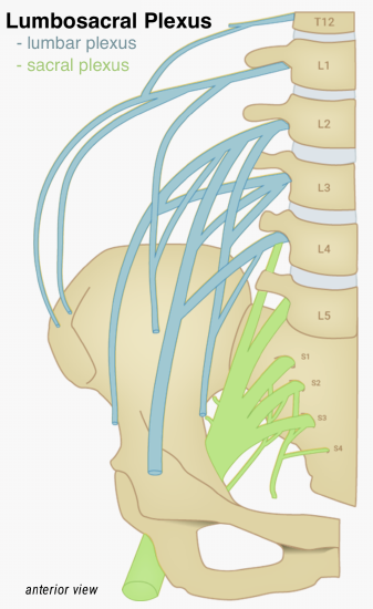
The lumbar plexus is the network of nerves that arises the anterior rami of lumbar spinal nerves L1 to L4, with a small contribution from T12. The nerves arising from this group innervate the inferior abdominal wall, genitalia, and the anteromedial thigh and leg. The sacral plexus arises from the anterior rami of spinal nerves L4-S4. Nerves from the sacral plexus innervate the gluteal region, pelvis, perineum, posterior thigh and most of the leg and foot. The two plexuses are sometimes grouped together into the lumbosacral plexus, since there is overlap at the L4 level (Figure \(\PageIndex{7}\)).
Named Nerves
Upper Limb - Branches from Brachial Plexus
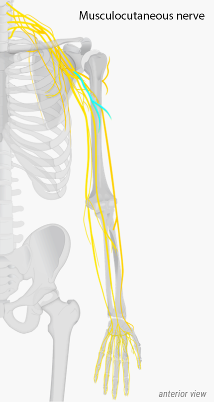
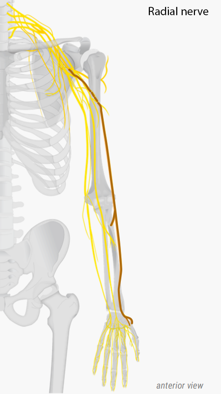
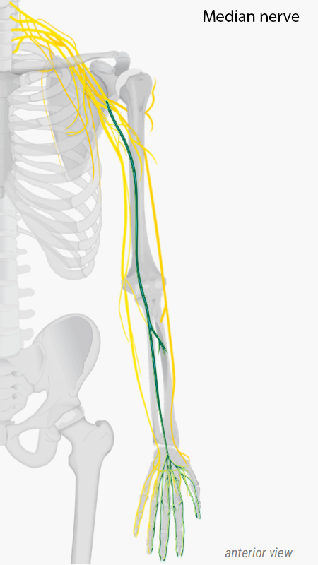
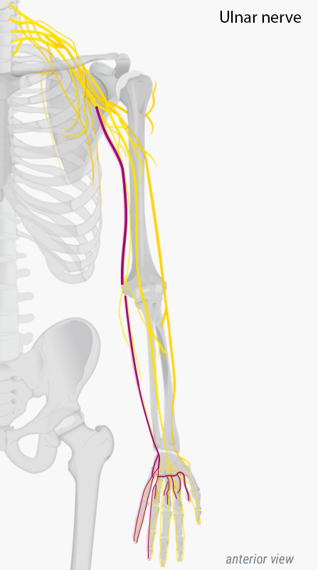
The brachial plexus gives rise to five terminal branches that innervate the upper limb: the axillary, median, musculocutaneous, radial and ulnar nerves (Figure \(\PageIndex{8}\)).
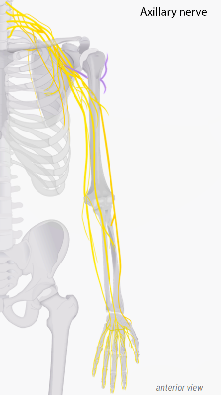
- axillary nerve innervates the deltoid and teres minor muscles, and receives sensation from the superolateral side of the arm
- median nerve innervates the most anterior forearm muscles as well as muscles that move the thumb. It receives sensation from the palmar region and dorsal tips of the lateral 3 and a half digits.
- musculocutaneous nerve innervate the anterior arm muscles and receives sensation from the lateral region of the forearm.
- radial nerve innervates muscles and receives sensation from the posterior arm and forearm, including the dorsal region of the lateral three digits (not including their tips).
- ulnar nerve innervates anterior forearm muscles as well as intrinsic hand muscles. It receives sensation from the dorsal and palmar regions of the medial one and a half digits.
Lower Limb - Branches from the Lumbar and Sacral Plexi
The lumbosacral plexus has two main branches that then give rise to other named nerves as well as several smaller branches. All innervate the lower limb and the pelvic region (Figure \(\PageIndex{9}\) and Figure \(\PageIndex{10}\)).
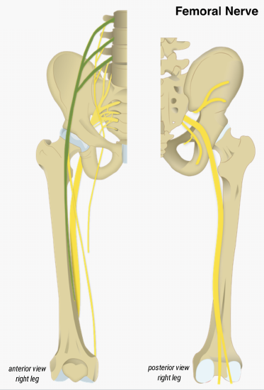
- femoral nerve: the main branch from the lumbar plexus; its small branches innervate anterior thigh muscles and receives sensory information from the anterior and medial aspects of the thigh.
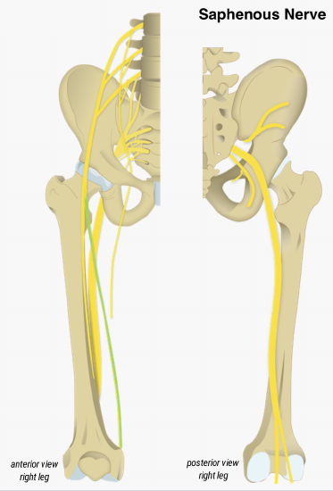
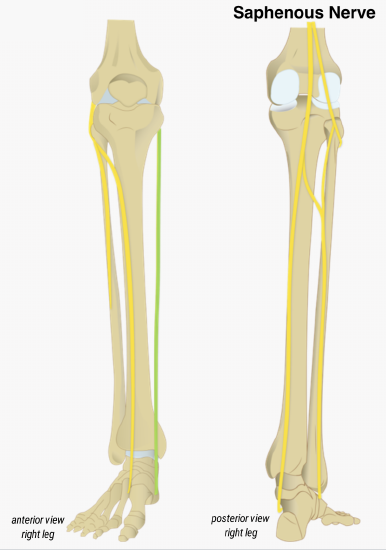
- saphenous nerve: a large branch of the femoral nerve that provides sensation from the medial aspect of the lower leg and the foot. It is the longest nerve in the body.
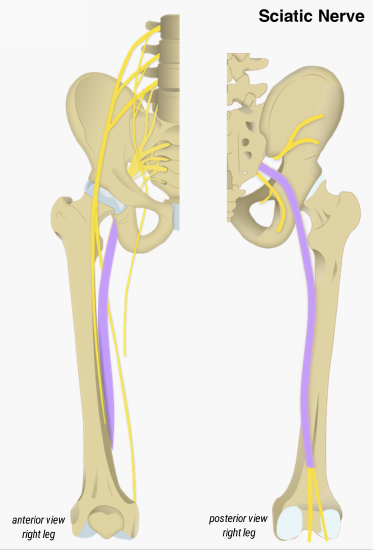
- sciatic nerve: the main branch from the sacral plexus and the largest and longest nerve of the body. It extends posteriorly across the hip joint and motor and sensory fibers to the posterior thigh and lower leg. The sciatic nerve bundles together two other nerves:
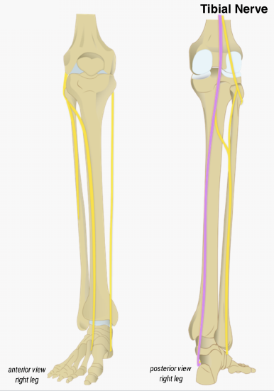
- tibial nerve: branch of the sciatic nerve that runs posteriorly along the midline of the lower limb and innervates the hamstrings, the posterior leg muscles, plantar foot muscles, and skin of the plantar foot.
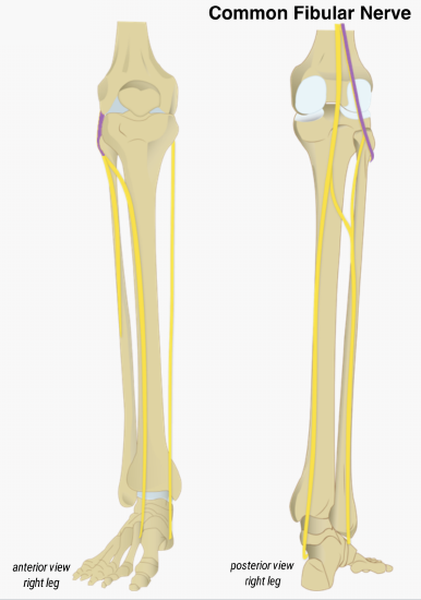
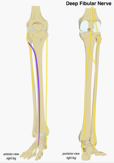
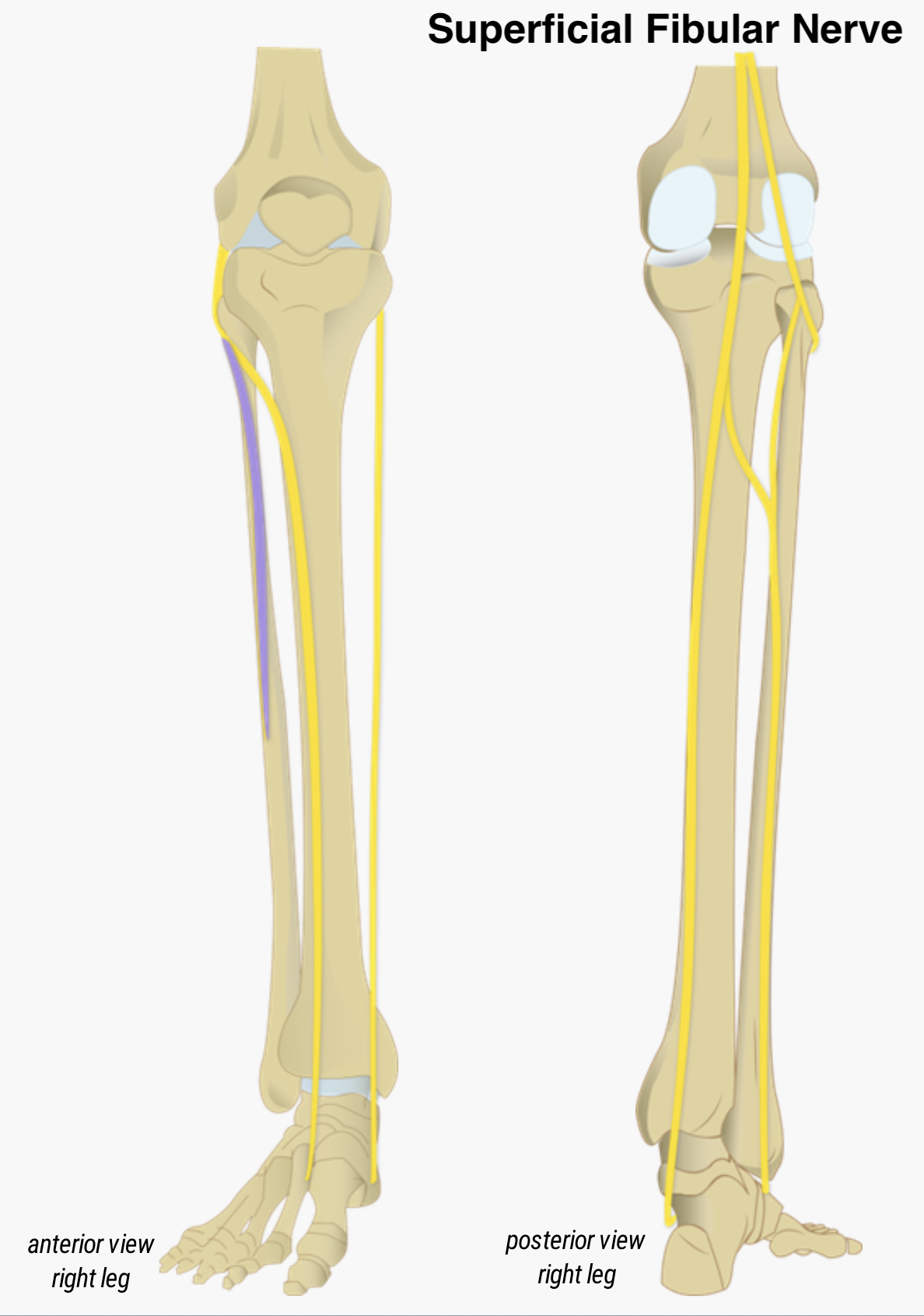
- common fibular nerve: branch of the sciatic nerve that wraps around the lateral knee and splits into two main branches:
- deep fibular nerve: runs in the anterior compartment of the leg to innervate the muscles of that compartment and the skin between the first and second toes.
- superficial fibular nerve: runs in the lateral compartment of the leg to innervate the muscles of that compartment as well as the muscles and skin on the superior surface of the foot.
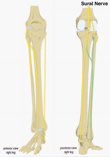
- sural nerve: arises in the midline sural (calf) region with fibers from both the common fibular and tibial nerves. It is a purely sensory nerve serving the posterior and lateral leg, ankle, and foot.
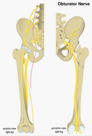
- obturator nerve: arises from the lumbar plexus and provides motor and sensory innervation to the medial thigh.
DISORDERS OF THE...
Sciatic Nerve
The sciatic nerve is most commonly associated with the painful condition sciatica, which is the result of compression or irritation of the nerve itself or of any of the spinal nerves giving rise to it.
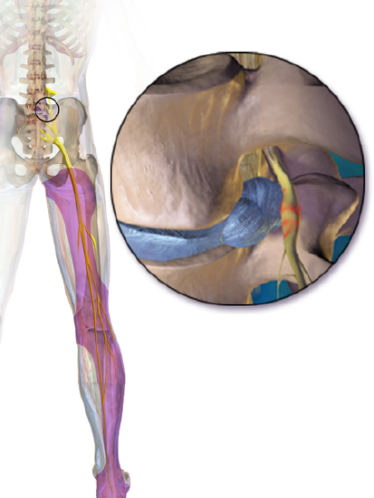
Sciatica most often occurs when either a herniated disc or an overgrowth of bone puts pressure on part of the nerve (Figure \(\PageIndex{11}\)) causing inflammation, pain, and, sometimes, numbness. Pain and numbness occur only on the affected side and most likely travel along the course of the nerve, from the gluteal region through the posterior thigh and into the posterolateral calf and foot. Pain can range from a mild ache to a sharp burning pain or can feel like a bolt of electricity. Numbness can range from a mild tingling (similar to when your leg falls asleep) to a lack of sensation in the area. Severe sciatica can also result in muscle weakness in the leg or foot.
Sciatica can occur from a traumatic injury, but more likely it arises from chronic risk factors including: advanced age, obesity, prolonged sitting, diabetes, and excessive/improper movements (twisting, carrying heavy objects, lifting, etc.).
Dermatomes
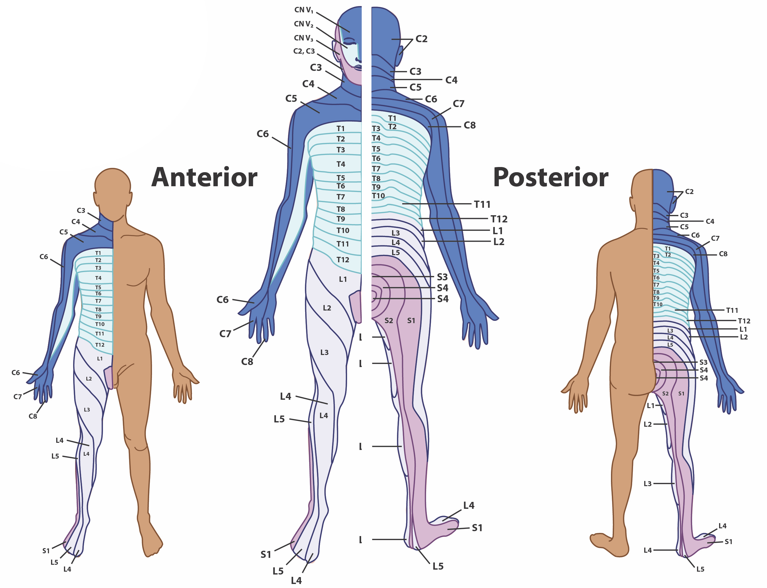
The spinal nerves, which contain sensory fibers with endings in the skin, connect with the skin in a topographically organized manner, illustrated as dermatomes (Figure \(\PageIndex{12}\)). For example, the fibers of eighth cervical nerve innervate the medial surface of the forearm and extend out to the fingers. All spinal nerves except C1 innervates a region on the skin. Dermatomes are important in a clinical setting as they can help identify potential damage to spinal nerves. In a sensory exam, the dermatome of a specific area is tested for sensation (for example the stroking of a cotton ball) to evaluate the functioning of the spinal nerve connected to that dermatome. Dermatomes are also associated with referred visceral pain, as the spinal nerves innervated the regions of the skin also innervate internal organs. Pain from the internal organ can then be mistakenly referred to a dermatome. For example, myocardial infarction is perceived as referred pain in the chest, left shoulder and left arm. This is because the sensory fibers that innervate the cardiac tissue travel through the T1-T4 spinal nerves, which also innervate the medial and anterior side of the body.
Concept Review
The spinal cord is covered and protected by the spinal meninges, similar to the cranial ones. However, the spinal dura mater presents only one layer of tissue. Moreover, the epidural space is a real space that houses areolar and adipose connective tissues, and blood vessels.
The spinal cord is divided into segments that take the same name as the vertebral regions. However, the sacral spinal cord ends at the vertebral lumbar level (L1/L2) with the conus medullaris, while the spinal nerves continue exiting at their respective region of the vertebral column. Inferior to the conus medullaris, the spinal nerves form a bundle called the cauda equina. The spinal cord presents two enlargements at the cervical and lumbar level.
In cross section, the gray matter is deep within the spinal cord and is separated into horns. The sensory axons coming from the posterior root ganglia enter the spinal cord through the posterior nerve root and synapse in the posterior horn, where interneurons are found. Autonomic motor neurons are found in the lateral horns, while somatic motor neurons are found in the anterior horns. Motor neurons send their axons out of the spinal cord through the anterior nerve root. The posterior and anterior root fuse to form the spinal nerve, which contains mixed sensory and motor information. The white matter is separated into columns which contain axons of sensory and motor neurons. The posterior columns contain ascending sensory tracts which are bundles of axons going towards the brain. The lateral and anterior column contain ascending sensory tracts as well as descending motor tracts, which carry the motor responses from the brain. Overall, the posterior regions of the spinal cord are responsible for ascending sensory information, while the anterior regions are responsible for descending motor information.
Spinal nerves are all mixed nerves with both sensory and motor fibers. Spinal nerves emerge from the spinal cord and reorganize through plexuses, which then give rise to systemic nerves. Thoracic spinal nerves are not part of any plexus, but give rise to the intercostal nerves directly.
Review Questions
Q. Which major nerve arises from the brachial plexus?
A. phrenic nerve
B. musculocutaneous nerve
C. intercostal nerve
D. saphenous nerve
- Answer
-
B
Q. Which regions of the body are innervated by the posterior rami?
A. head and neck
B. upper limbs
C. posterior thoracic and lumbar
D. lower limbs
- Answer
-
C
Q. The anterior root carries which type of information?
A. motor only
B. sensory only
C. both motor and sensory
- Answer
-
A
Glossary
- axillary nerve
- systemic nerve of the arm that arises from the brachial plexus
- brachial plexus
- nerve plexus associated with the lower cervical spinal nerves and first thoracic spinal nerve
- cervical plexus
- nerve plexus associated with the upper cervical spinal nerves
- cord
- branch of the brachial plexus originating from the divisions
- dermatome
- topographical area of skin in which sensory nerves end
- dorsal (posterior) nerve root
- axons entering the posterior horn of the spinal cord
- dorsal ramus
- dorsal branch of a spinal nerve that forms from the dorsal root of the nerve after it emerges from the spinal cord
- dorsal root ganglion
- cluster of cell bodies in the dorsal root of a spinal nerve containing sensory neurons
- femoral nerve
- systemic nerve of the anterior leg that arises from the lumbar plexus
- fibular nerve
- systemic nerve of the posterior leg that begins as part of the sciatic nerve
- intercostal nerve
- systemic nerve in the thoracic cavity that is found between two ribs
- lumbar plexus
- nerve plexus associated with the lumbar spinal nerves
- median nerve
- systemic nerve of the arm, located between the ulnar and radial nerves
- musculocutaneous nerve
- systemic nerve of the arm, located lateral to the other nerves of the brachial plexus
- nerve plexus
- network of nerves without neuronal cell bodies included
- phrenic nerve
- systemic nerve from the cervical plexus that innervates the diaphragm
- radial nerve
- systemic nerve of the arm, the distal component of which is located near the radial bone
- ramus
- branch of a spinal nerve
- rami communicans (communicantes)
- structures that provide a short connection from an autonomic ganglion to the spinal nerve
- sacral plexus
- nerve plexus associated with the lower lumbar and sacral spinal nerves
- saphenous nerve
- systemic nerve of the lower anterior leg that is a branch from the femoral nerve
- sciatic nerve
- systemic nerve from the sacral plexus that is a combination of the tibial and fibular nerves and extends across the hip joint and gluteal region into the upper posterior leg
- sciatica
- painful condition resulting from inflammation or compression of the sciatic nerve or any of the spinal nerves that contribute to it
- spinal nerve
- one of 31 nerves connected to the spinal cord
- systemic nerve
- nerve in the periphery distal to a nerve plexus or spinal nerve
- tibial nerve
- systemic nerve of the posterior leg that begins as part of the sciatic nerve
- ulnar nerve
- systemic nerve of the arm located close to the ulna, a bone of the forearm
- ventral (anterior) nerve root
- axons emerging from the anterior or lateral horns of the spinal cord
- ventral ramus
- ventral branch of a spinal nerve that forms from the ventral root of the nerve after it emerges from the spinal cord
Contributors and Attributions
OpenStax Anatomy & Physiology (CC BY 4.0). Access for free at https://openstax.org/books/anatomy-and-physiology